The Brain on the Wall: Robert E. Holding, E.J. Arnold & Son, and the Edwardian Anatomy Chart
Before the projector, before the digital whiteboard, before the internet made every anatomical image instantly accessible, the science classroom had the wall chart. Large, detailed, precisely illustrated, it hung at the front of the room where every student could see it — a permanent presence in the educational environment, a visual reference that shaped the way a generation understood the human body. The great anatomical wall charts of the late nineteenth and early twentieth centuries were not merely teaching aids. They were, in the fullest sense, works of art: images that combined scientific accuracy with visual clarity and aesthetic ambition, that made the invisible structures of the body visible and comprehensible to students who had never seen a dissection and never would.
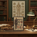
Robert E. Holding’s chart of the brain and nervous system, published by E.J. Arnold & Son Ltd. of London, Edinburgh, and Belfast, was one of the finest examples of this tradition. Designed for the British science classroom of the early twentieth century, it showed the complete anatomy of the human nervous system — the brain and cerebellum, the spinal cord, the cranial nerves, the peripheral nervous system — with a precision and clarity that made it both a reliable scientific reference and a visually compelling image. It is the kind of illustration that rewards sustained attention: the more carefully you look, the more you see.
E.J. Arnold & Son and the British Educational Publishing Tradition
To understand Robert E. Holding’s chart, it is necessary to understand the company that published it. E.J. Arnold & Son Ltd. was one of the leading educational publishers in Britain in the late nineteenth and early twentieth centuries. Founded in Leeds in 1863 by Edward John Arnold, the company specialised in educational materials — textbooks, exercise books, maps, charts, and the full range of printed materials that the expanding British school system required. By the early twentieth century, it had offices in London, Edinburgh, and Belfast, and its products were used in schools throughout the United Kingdom and the British Empire.
The company’s wall charts were among its most important products. Designed to be displayed in classrooms, they covered a wide range of subjects — geography, natural history, history, and science — and were produced to a consistently high standard of illustration and printing. The science charts, in particular, were notable for their combination of accuracy and visual clarity: they were designed to be understood by students who were encountering the subject for the first time, but they were also accurate enough to serve as genuine scientific references.
The tradition of the educational wall chart that E.J. Arnold & Son represented had its roots in the natural history illustrations of the eighteenth century, but it reached its fullest development in the second half of the nineteenth century, when improvements in printing technology — particularly chromolithography — made it possible to produce large-format, high-quality images at relatively low cost. By the early twentieth century, the wall chart had become a standard feature of the British classroom, and publishers like E.J. Arnold & Son were producing series of charts covering every subject in the school curriculum.
Reading the Brain: Holding’s Chart and the Anatomy of the Nervous System
The subject of Holding’s chart — the brain and nervous system — was one of the most challenging in the science curriculum. The nervous system is, by any measure, the most complex system in the human body: a network of billions of cells, connected by trillions of synapses, responsible for every sensation, every movement, every thought and feeling and memory. To represent this system in a single image, in a way that was both accurate and comprehensible to a student encountering it for the first time, required exceptional skill in both scientific knowledge and visual communication.
Holding’s chart met this challenge with considerable success. The central image shows the brain in lateral view — the cerebral cortex with its characteristic folds and fissures, the cerebellum at the rear, the brainstem connecting the brain to the spinal cord. Surrounding this central image are detailed diagrams of the component structures: the cranial nerves, shown in their relationship to the brain and the structures they innervate; the spinal cord in cross-section, showing the arrangement of grey and white matter; the peripheral nervous system, showing how the spinal nerves branch out to supply the muscles and skin of the body.
The labeling is precise and systematic: every structure is named, every relationship is indicated. The color coding — a standard feature of anatomical illustration by the early twentieth century — helps to distinguish different types of tissue and different functional systems. The overall composition is carefully designed to guide the eye through the complexity of the subject, from the central image of the brain to the surrounding detail diagrams, in a sequence that builds understanding progressively.
It is, in short, a masterwork of scientific illustration — an image that does its work so well that it is easy to overlook the skill required to produce it.
The Human Body and Health: The Textbook Tradition
The back cover of our journal carries an illustration from a different but related tradition: The Human Body and Health — An Elementary Text-Book of Essential Anatomy, Applied Physiology and Practical Hygiene for Schools, published in 1908. Where Holding’s chart was designed for display — to hang on the wall and be consulted repeatedly over the course of a school year — the textbook illustration was designed to be studied in the hand, to accompany a text that explained and elaborated on what the image showed.
The textbook tradition of anatomical illustration had a long history by 1908. Andreas Vesalius’s De Humani Corporis Fabrica of 1543 — with its extraordinary woodcut illustrations of the human body — had established the standard for anatomical illustration that subsequent centuries would refine but not fundamentally alter: the combination of precise observation, skilled draughtsmanship, and systematic labeling that made the image both scientifically accurate and visually comprehensible.
By 1908, the tradition had been enriched by three and a half centuries of anatomical research and illustrative practice. The illustrations in The Human Body and Health reflect this accumulated knowledge: they show structures that Vesalius could not have known, rendered with a precision that his woodcut technique could not have achieved, in a format designed for the mass educational market that his aristocratic patrons could not have imagined. They are, in their way, as remarkable as their sixteenth-century predecessors — not for their novelty, but for the quality of their synthesis: the ability to distil centuries of anatomical knowledge into images that a schoolchild could understand.
The Edwardian Science Classroom
The charts and textbooks that Holding illustrated and E.J. Arnold & Son published were designed for a specific educational environment: the Edwardian science classroom. By the early twentieth century, science had become a standard part of the British school curriculum — a development that would have seemed radical to the educators of a generation earlier, for whom the classics and mathematics were the core of a proper education.
The inclusion of science in the curriculum reflected a broader shift in British culture: a growing recognition that the industrial and commercial success of the nation depended on a workforce with scientific and technical knowledge, and that the schools had a responsibility to provide that knowledge. The Education Acts of the late nineteenth century had established a system of compulsory elementary education, and the science curriculum that developed within this system was designed to give every child a basic understanding of the natural world and the human body.
Physiology and hygiene — the study of how the body works and how to keep it healthy — were particularly important parts of this curriculum. In an era before antibiotics, before effective vaccines for most infectious diseases, before the germ theory of disease had fully penetrated popular understanding, the teaching of basic hygiene was a matter of genuine public health significance. The anatomical charts and physiology textbooks of the Edwardian period were not merely educational materials: they were instruments of public health, designed to teach a generation of children the basic principles of bodily function and personal hygiene that could, quite literally, save lives.
Anatomical Illustration as Art
There is a tendency to think of anatomical illustration as purely functional — as a form of scientific documentation rather than artistic expression. This tendency does a disservice to the illustrators who produced these images. The great anatomical illustrators of the nineteenth and early twentieth centuries — among whom Robert E. Holding deserves to be counted — were artists of considerable skill and sensitivity, working in a tradition that demanded both scientific accuracy and visual elegance.
The challenge of anatomical illustration is, in some ways, more demanding than that of other forms of scientific illustration. The natural history illustrator can observe a living specimen and render what they see; the anatomical illustrator must represent structures that are, in the living body, invisible — hidden beneath skin and muscle and bone, revealed only by dissection. The image must be accurate enough to serve as a scientific reference, clear enough to be understood by a student, and visually compelling enough to hold the attention of a classroom full of children who might otherwise find the subject intimidating.
Holding’s chart meets all of these demands. The brain, rendered in careful detail with its characteristic folds and fissures, is immediately recognisable as a brain — but it is also, in the quality of its rendering, a beautiful object. The color coding that distinguishes different structures is not merely functional: it gives the image a visual richness that makes it satisfying to look at quite apart from its informational content. The overall composition — the central image surrounded by detail diagrams, the careful arrangement of labels and annotations — has a visual coherence that reflects genuine aesthetic judgment.
These are images that reward sustained attention — that reveal more the longer you look at them. They belong to a tradition of scientific illustration that understood that beauty and accuracy are not opposites, that an image can be both scientifically rigorous and visually compelling, that the best way to teach is also, often, the most beautiful.
A Journal for Those Who Study the Mind

Our Brain & Nervous System Anatomy Journal carries these two 1908 illustrations across its full wraparound cover — Holding’s brain chart on the front, the Human Body and Health illustration on the back. It is a journal for those who find beauty in anatomical illustration, who understand that a diagram of the nervous system is also a kind of portrait of what it means to be human, who appreciate the Edwardian tradition of scientific education that produced these images.
Inside, 150 perforated lined pages await your medical notes, neuroscience observations, anatomy studies, or whatever form your engagement with the science of the mind takes. The casewrap sewn binding opens completely flat — ideal for sketching anatomical diagrams. The matte laminated cover preserves every detail of Holding’s illustration in a finish that rewards close examination.
In 1908, a generation of British schoolchildren learned about the brain from a chart on the wall. Perhaps the pages inside will help you understand it a little better too.
References & Further Reading
- Cunningham, Andrew & Williams, Perry (eds.). The Laboratory Revolution in Medicine. Cambridge University Press, 1992.
- Kemp, Martin. The Human Animal in Western Art and Science. University of Chicago Press, 2007.
- Laqueur, Thomas. Making Sex: Body and Gender from the Greeks to Freud. Harvard University Press, 1990.
- Netter, Frank H. Atlas of Human Anatomy. Elsevier, 1989.
- Richardson, Ruth. The Making of Mr Gray’s Anatomy. Oxford University Press, 2008.
- Vesalius, Andreas. De Humani Corporis Fabrica. Basel, 1543.
- Warner, John Harley & Edmonson, James M. Dissection: Photographs of a Rite of Passage in American Medicine 1880–1930. Blast Books, 2009.