The Body as Atlas: Larousse Médical Illustré and the Art of French Anatomical Illustration
In 1912, to open the Larousse Médical Illustré was to enter a world in which the human body had been made legible — its systems mapped, its structures named, its extraordinary complexity rendered visible in diagrams of such precision and clarity that the reader could follow the branching of a nerve from the brain to the fingertip, or trace the arrangement of the abdominal organs from the diaphragm to the pelvis. It was, in the fullest sense of the word, an atlas of the self.
The Larousse Médical Illustré was published in Paris in 1912, edited by Dr. Galtier-Boissière, and issued by the Librairie Larousse — the publishing house that had been producing encyclopedias, dictionaries, and reference works for the French public since Pierre Larousse founded it in 1852. By 1912, the Larousse name was synonymous with authoritative, accessible knowledge: the Grand Dictionnaire Universel du XIXe Siècle, the Nouveau Larousse Illustré, and a succession of specialised encyclopedias had established the house as the preeminent publisher of popular reference works in France. The Médical Illustré was conceived in the same spirit: a comprehensive guide to medicine and human anatomy, written for the educated general reader rather than the specialist, and illustrated with a thoroughness and quality that set it apart from anything previously available to a non-professional audience.
The timing was significant. French medicine in 1912 was at a moment of extraordinary productivity. The germ theory of disease, established by Pasteur and Koch in the previous decades, had transformed the understanding of infection and immunity. The discovery of X-rays by Röntgen in 1895 had opened new possibilities for the visualisation of the body’s interior. The development of anaesthesia and antiseptic technique had made surgery safer and more ambitious. And the systematic study of neuroanatomy — the structure and function of the brain and nervous system — was advancing rapidly, driven by the work of figures like Cajal, Golgi, and the French neurologist Jean-Martin Charcot. The Larousse Médical Illustré appeared at the moment when all of this knowledge was ready to be synthesised and communicated to a wider public.
The Plates: Brain, Nerves, and Viscera
The anatomical illustrations in the Larousse Médical Illustré are among the finest examples of medical art produced in the early twentieth century. Rendered against a black background — a technique borrowed from the natural history illustration tradition, where it had been used to isolate specimens with maximum clarity — they combine the precision of scientific draughtsmanship with a graphic intensity that makes them immediately striking as visual objects.
Planche IX, page 191 — "Cerveau Nerfs Partant du" (Brain and Departing Nerves) — shows the human brain in lateral view, with the cranial nerves emerging from its base and fanning out toward their destinations in the face, the neck, and the body. The labeling is meticulous: each nerve is identified by name and number, each structure of the brain surface named and located. The color is used functionally — different systems distinguished by different hues — but the overall effect is also beautiful: the brain rendered in warm grey-pink against the black ground, the nerves in their various colors spreading outward like the roots of a tree.
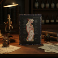
The viscera plate — "Viscères de l’abdomen et du thorax" — presents four views of the human torso, showing the arrangement of the thoracic and abdominal organs from different angles and at different levels of dissection. Heart, lungs, liver, stomach, intestines, kidneys, and the major blood vessels are all labeled with the precision that the medical student or the curious general reader would require. The four-view format — a convention of anatomical illustration since Vesalius — allows the reader to build up a three-dimensional understanding of the body’s interior from a series of two-dimensional images, each one adding a layer of information to the picture that the others have established.
Jacques Maurice Dessertenne and the Tradition of Medical Illustration
The illustrations in the Larousse Médical Illustré are attributed, in the scholarly literature, to Jacques Maurice Dessertenne — a French illustrator who worked extensively for the Larousse publishing house in the early twentieth century and whose work appeared in a number of their encyclopedic publications. Medical illustration in this period was a specialised discipline that required both artistic skill and scientific knowledge: the illustrator had to be able to work from anatomical specimens, from dissections, and from the existing scientific literature, producing images that were accurate enough to be useful to a medical professional and clear enough to be understood by a general reader.
Dessertenne’s work in the Larousse Médical Illustré meets both requirements. His illustrations are scientifically accurate — the structures they depict correspond to what the anatomist would find in the dissecting room — and they are also, in the best tradition of French scientific illustration, genuinely beautiful. The black background, the careful use of color to distinguish different systems, the precise labeling, and the confident draughtsmanship combine to produce images that reward both the student who needs to learn from them and the viewer who simply finds the human body extraordinary.
Medicine Made Legible
The Larousse Médical Illustré was part of a broader project of popular scientific education that characterised the Belle Époque in France — the belief, widespread among the educated classes, that scientific knowledge should be accessible to everyone, that the discoveries of the laboratory and the clinic should not remain the exclusive property of specialists but should be communicated, in clear and beautiful form, to the general public. It was a belief that produced, in the decades around 1900, some of the finest popular science writing and illustration ever made — works that took seriously both the complexity of their subject matter and the intelligence of their readers.
The anatomical plates of the Larousse Médical Illustré are among the lasting achievements of this tradition. They made the human body legible — not by simplifying it, but by rendering its complexity with sufficient clarity and beauty that the reader could engage with it directly, without the mediation of a specialist. More than a century later, they continue to do exactly that.

If the history of French medicine and the art of anatomical illustration resonate with you, the Larousse Médical Journal brings Dessertenne’s 1912 brain and viscera plates to a hardcover journal — 150 lined pages, ready for anatomy notes, clinical observations, or whatever your studies require.
References
- Galtier-Boissière, Dr. (ed.) Larousse Médical Illustré. Librairie Larousse, Paris, 1912.
- Cazort, M., Kornell, M. & Roberts, K.B. The Ingenious Machine of Nature: Four Centuries of Art and Anatomy. National Gallery of Canada, Ottawa, 1996.
- Rifkin, B.A., Ackerman, M.J. & Folkenberg, J. Human Anatomy: Depicting the Body from the Renaissance to Today. Thames & Hudson, London, 2006.
- Ackerknecht, E.H. Medicine at the Paris Hospital, 1794–1848. Johns Hopkins Press, Baltimore, 1967.
- Sournia, J.-C. A History of Medicine. Blackwell, Oxford, 1992.