The Body Mapped: Gustave H. Michel and the American Anatomical Chart of 1910
Share
In the early years of the twentieth century, the American medical school was undergoing a transformation. The Flexner Report of 1910 — the landmark survey of medical education commissioned by the Carnegie Foundation — would soon reshape the entire landscape of medical training in North America, closing dozens of inadequate schools and establishing rigorous standards for the rest. But before Flexner, and in the same year his report appeared, another kind of document was being produced in Cleveland, Ohio: the anatomical charts of Dr. Gustave H. Michel.
Published by Dr. G.H. Michel & Co. and copyrighted in both Europe and America in 1910, Michel's charts were designed for exactly the kind of medical education that Flexner was trying to improve — the teaching of anatomy through visual documentation, the making-visible of systems that no student could see without dissection. They were, in the fullest sense, teaching tools: large-format lithographic scroll charts, measuring approximately 48 × 37 inches, designed to hang on the walls of medical schools and preparation rooms and to be consulted, pointed at, and studied by generations of students.
The Chart of the Arterial, Venous and Nervous Systems
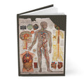
The front cover of this journal reproduces Michel's Chart of the Arterial, Venous and Nervous Systems — a highly detailed lithographic document illustrating the major circulatory and nervous systems of the human body. The central figure presents the complete arterial and venous network from head to foot, with color-coded vessels tracing the pathways of blood through the body's architecture. Surrounding it: specific anatomical views of the brain showing neural pathways, the heart displaying its chambers and major vessels, and internal organs with their vascular and nervous connections.
The level of detail is extraordinary. Every named artery, every major nerve, every organ's blood supply is documented with the precision that a medical student would require — and with the graphic confidence of a lithographer working at the height of his craft.
The Chart of the Muscular System
The back cover presents Michel's Chart of the Muscular System — comprehensive anterior and posterior views of both superficial and deep muscle layers, allowing students to understand complete muscular anatomy from multiple perspectives. Detailed inserts include specific diagrams of the head and neck musculature, intricate hand and foot muscle groups, and a cross-section of the head revealing the brain and nasal cavity structures.
Together, the two charts form a complete atlas of the human body's active systems — the networks that carry blood and nerve impulses, and the muscles that move the skeleton through the world.
Cleveland and the Medical Publishing Tradition
That these charts were produced in Cleveland is not incidental. By 1910, Cleveland was one of America's great medical cities — home to Western Reserve University's medical school, to the Cleveland Clinic (founded 1921), and to a tradition of medical publishing that reflected the city's ambitions as a center of scientific education. Dr. G.H. Michel & Co. was part of this tradition: a publishing house that understood the visual demands of medical education and had the lithographic resources to meet them.
The decision to copyright the charts in both Europe and America speaks to their ambition. These were not local teaching aids but documents intended for an international market — for the medical schools of Germany and Britain as much as for those of Ohio and New York.
Explore the journal: