Cœur et Nerfs: How French Medical Publishing Taught a Nation to See the Human Body
I. The Problem of the Invisible Body
For most of human history, the interior of the body was inaccessible — not only physically, but visually. Anatomy was a discipline of specialists, conducted in dissection theatres before small audiences of medical students, its findings recorded in texts that circulated among the learned and nowhere else. The body’s systems — the branching networks of arteries and veins, the intricate architecture of the nervous system, the chambers of the heart — existed, for most people, as abstractions: named but unseen, described but unimaginable.
The problem was not merely one of access. It was one of representation. Even for those with access to anatomical texts, the illustrations available before the mid-nineteenth century were often schematic, inconsistent, and difficult to interpret. The gap between the written description and the visual reality of the body was enormous, and it was a gap that had real consequences for medical education.
The chromolithographic revolution of the second half of the nineteenth century began to close that gap. And in France, no institution closed it more completely — or more beautifully — than the house of Larousse.
II. Pierre Larousse and the Democratic Encyclopedia
Pierre Larousse (1817–1875) was not a physician. He was a lexicographer, a pedagogue, and a man of the French republican left who believed, with the conviction of his generation, that knowledge was a political instrument — that to make information accessible to ordinary people was to make them free.
His great project, the Grand dictionnaire universel du XIXe siècle, published between 1866 and 1876, was conceived as an encyclopedia for everyone: not for the scholar in his library, but for the schoolteacher in a provincial town, the artisan in his workshop, the citizen who wanted to understand the world he lived in. It was illustrated, affordable by the standards of the time, and written in a French that did not require a university education to read.
The medical volumes that followed — the Dictionnaire complet illustré and, later, the Larousse Médical Illustré — carried the same democratic ambition into the domain of the body. The human body, Larousse’s successors argued, was not the exclusive property of the medical profession. Every person had a body; every person had a right to understand it.
III. Galtier-Boissière and the Larousse Médical
Jean Galtier-Boissière (1876–1953) was the physician and editor who brought this ambition to its fullest realization. A graduate of the Paris Faculty of Medicine, he was appointed director of the Larousse Médical Illustré in the early years of the twentieth century, and the edition he produced — published in 1912 — became one of the most widely distributed medical reference works in French history.
The Larousse Médical Illustré of 1912 was not a textbook for specialists. It was a reference work for the educated general public: for the family that wanted to understand a diagnosis, for the nurse who needed a visual reference, for the student who was encountering the body’s systems for the first time. Its language was precise but accessible; its illustrations were designed to be understood at a glance.
Galtier-Boissière understood that the illustration was not a supplement to the text — it was, for many readers, the primary means of understanding. He commissioned plates of extraordinary quality, working with artists trained in both scientific accuracy and the chromolithographic techniques that had, by 1912, reached their highest point of refinement.
IV. The Chromolithographic Tradition
Chromolithography — the process of printing in multiple colors from a series of lithographic stones, each carrying a different color — had been developed in the 1830s and 1840s, and by the second half of the nineteenth century it had transformed the production of illustrated books and periodicals across Europe.
For medical illustration, chromolithography offered something that no previous technique could provide: the ability to reproduce, in full color and at scale, the complex visual information of the human body. The red of arterial blood, the blue of venous return, the grey of nervous tissue, the yellow of fat and connective tissue — all of these could now be rendered with a fidelity that black-and-white engraving could not approach.
The best chromolithographic medical plates of the period are works of considerable artistic achievement. They required not only scientific accuracy — the illustrator had to know anatomy — but also a mastery of color, composition, and the specific technical demands of the lithographic process. The plates were produced by a collaboration between the physician-editor, who specified the content, and the artist, who translated that content into visual form.
In the case of the Larousse Médical Illustré, that artist was Leuba — whose full name has not survived in the historical record, but whose work has. Leuba’s plates were executed first as watercolors, with the precision and delicacy that the medium demands, and then transferred to the lithographic stone for reproduction. The result was a series of images that combined scientific rigor with genuine aesthetic refinement.
V. Cœur et Circulation — The Cardiovascular Plate
The plate Cœur et circulation (artères, veines, lymphatiques) — from the Dictionnaire complet illustré of Larousse, produced in the period between 1889 and 1912 — is a comprehensive map of the body’s circulatory systems. It traces the arterial network from the aorta through its major branches to the extremities; it shows the venous return; it maps the lymphatic vessels and nodes that run alongside the vascular system.
Supporting diagrams show the heart in cross-section — Coupe du cœur — with its four chambers and the valves that regulate the flow of blood. Microscopic diagrams show red and white blood cells — Globules rouges et blancs — at a scale that makes their form visible. The lymphatic valve structure is shown in detail.
The plate is, in effect, a complete visual account of the body’s transport systems — the networks that carry oxygen, nutrients, immune cells, and waste products through the body. It was designed to be read as a whole, the eye moving from the large-scale map of the body to the microscopic detail of the cell, understanding the system at every scale simultaneously.
VI. Cerveau et Nerfs — The Nervous System Plate
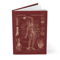
The plate Cerveau et Nerfs, Plate IX of the Larousse Médical Illustré of 1912, presents the human nervous system in the écorché tradition — the tradition of the flayed figure, in which the skin is removed to reveal the structures beneath.
The écorché had been a staple of anatomical illustration since the Renaissance, when artists and anatomists collaborated to produce images that showed the body’s musculature and skeletal structure in a way that no clothed or intact figure could. By the nineteenth century, the tradition had been extended to the nervous system — a subject that presented particular challenges, since the nerves are fine, branching structures that are difficult to show clearly in a single image.
Leuba’s solution was to show the nervous system in two views — anterior and posterior — with the body in a standing position and the nerves traced from the brain and spinal cord to their peripheral terminations. The anterior view shows the facial nerves, the plexus brachial that serves the arm, and the nerfs intercostaux that run between the ribs. The posterior view traces the system from the cerveau and cervelet down the moelle épinière to the nerf sciatique — the sciatic nerve, the longest in the body.
The French anatomical labels — cerveau, cervelet, moelle épinière, plexus brachial, nerf sciatique — are not merely decorative. They are part of the plate’s pedagogical function: to teach the names and locations of the nervous system’s major structures simultaneously, so that the student who has looked at the plate has learned both the anatomy and the vocabulary.
VII. The Inversion
The plates in their original form were printed in the chromolithographic palette of the period: warm flesh tones, the red of arteries, the blue of veins, the grey of nervous tissue, against a cream or white ground.
The versions that appear on this journal have been artistically inverted — the clinical black ink of the original line work transformed into warm lacre red against cream lines. The inversion is not a distortion of the original; it is a reinterpretation in the chromolithographic spirit, honoring the tradition of color in medical illustration while creating an aesthetic that belongs to the present as much as to 1912.
The result is an image that reads simultaneously as historical document and contemporary design — which is, perhaps, the most honest way to describe what these plates have always been.
VIII. The Legacy of the Larousse Médical
The Larousse Médical Illustré of 1912 went through numerous editions and remained in print for decades. It was the medical reference that French families kept on their shelves alongside the dictionary — the book you consulted when a diagnosis was unclear, when a symptom needed to be understood, when the body’s workings required explanation.
Its plates — Leuba’s chromolithographs of the cardiovascular and nervous systems, among others — were reproduced, copied, and adapted across the French-speaking world. They shaped the visual vocabulary through which a generation of students, nurses, and educated laypeople understood the human body.
That visual vocabulary has not entirely disappeared. The écorché figure, the color-coded vascular map, the labeled diagram of the nervous system — these are still the forms through which anatomy is taught, even if the medium has changed from chromolithography to digital rendering. Leuba’s plates are ancestors of every anatomical illustration produced since.
IX. A Note on This Journal

The plates on this journal are from the Larousse medical publishing tradition — Leuba’s chromolithographic illustrations of the cardiovascular and nervous systems, produced for Galtier-Boissière’s Larousse Médical Illustré of 1912 and the earlier Dictionnaire complet illustré. Artistically inverted into lacre red and cream, they carry the democratic ambition of Pierre Larousse into a new form: a hardcover journal for those who write the body in careful observation.
👉 Anatomy Journal — Larousse Médical 1912
References
- Galtier-Boissière, J. (ed.) (1912). Larousse Médical Illustré. Paris: Larousse.
- Larousse, P. (1866–1876). Grand dictionnaire universel du XIXe siècle. Paris: Larousse.
- Larousse (c.1889–1912). Dictionnaire complet illustré. Paris: Larousse.
- Rifkin, B.A., Ackerman, M.J. & Folkenberg, J. (2006). Human Anatomy: Depicting the Body from the Renaissance to Today. Thames & Hudson.
- Cazort, M., Kornell, M. & Roberts, K.B. (1996). The Ingenious Machine of Nature: Four Centuries of Art and Anatomy. National Gallery of Canada.
- Sappol, M. (2017). Hidden Treasure: The National Library of Medicine. Blast Books.